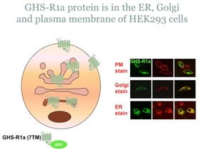
My study of GPCR dimerization started with the growth hormone secretagoue receptor-1a (GHS-R1a), later known as the ghrelin receptor after its endogenous agonist was finally identified. By mutating the gene sequence of GHS-R1a, I have added a green fluorescent protein (GFP) to the C-terminal tail which sits in the cytoplasm of the cell. By detecting the fluorescence emitted by GFP, one can 'see' the location of GHS-R1a in a cell which has been transfected with DNA capable of instructing the cell to make this receptor protein. By using cell stains which identify the ER and Golgi, one can then determine where GHS-R1a resides in a cell. As you can see in the figure below, GHS-R1a is found throughout the cell, but is clearly localized in the cell membrane. (To determine co-localization of proteins using fluorescence microscopy techniques, one can use a green signal for one protein and a red signal for the other protein. If the proteins are in a similar location, then you will see a yellow colour.)